Client: University of Wisconsin Radiology Department – Collaboration with an oncologist.
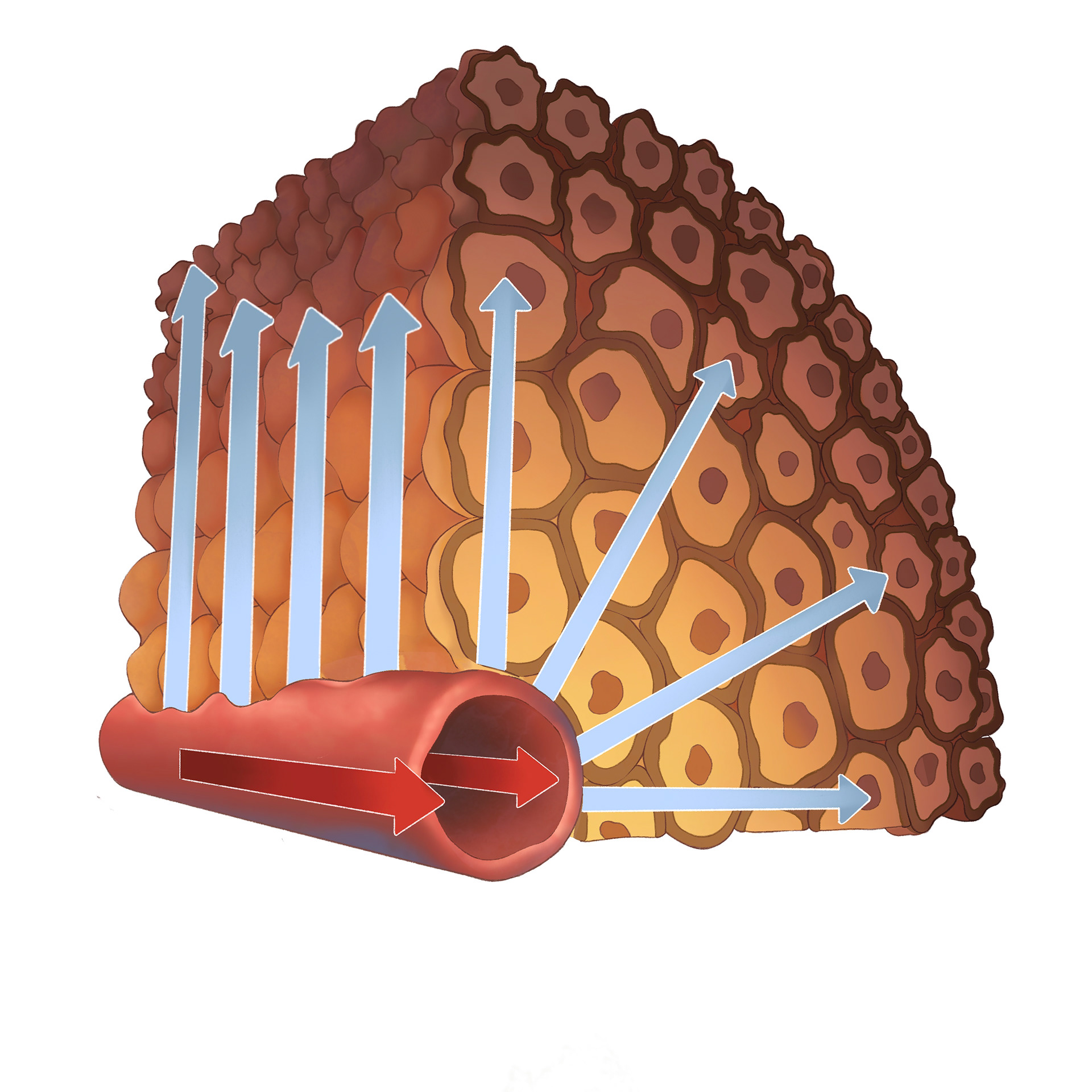
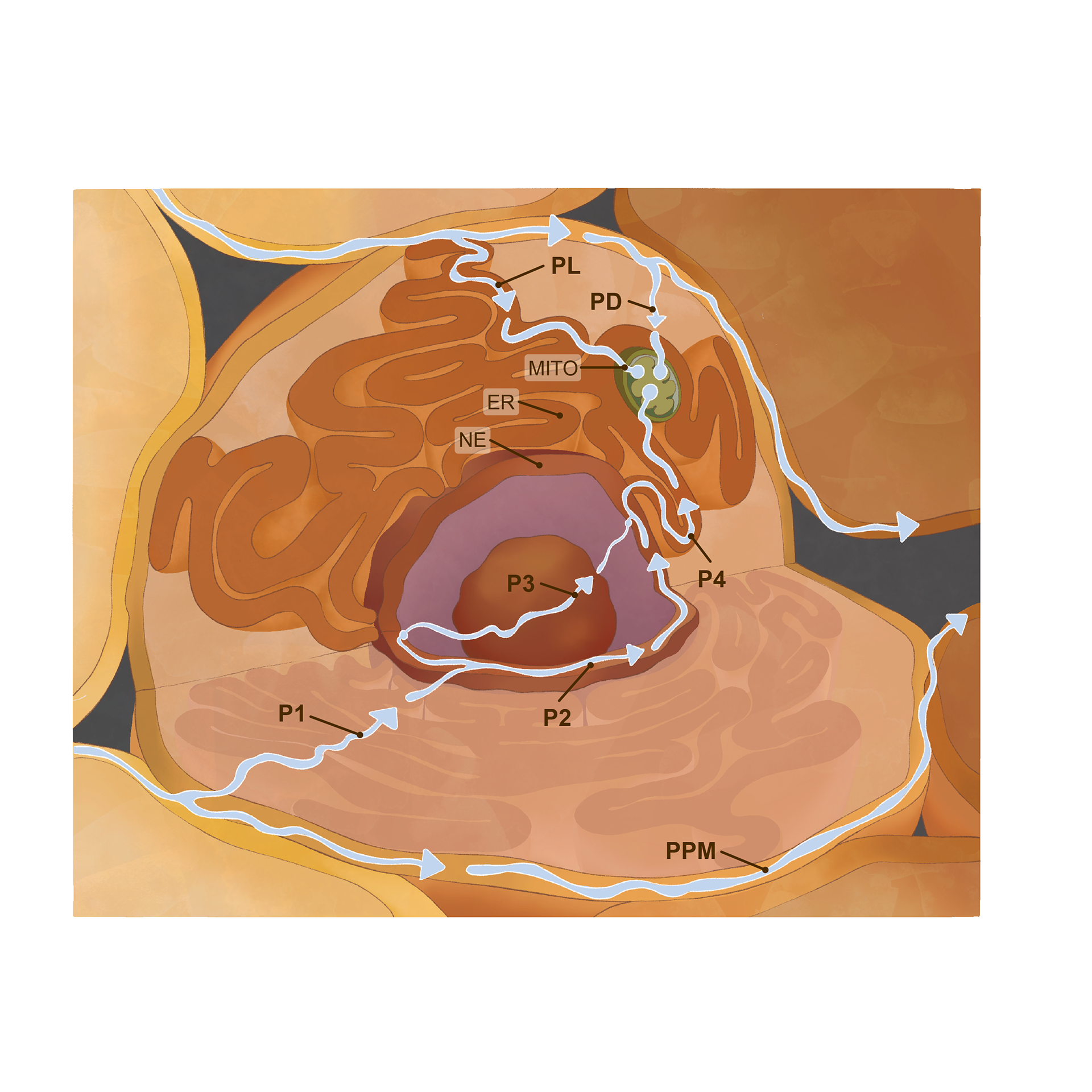
Purpose: Created a series of three scientific illustrations to visually communicate a new theoretical model of oxygen dissipation through cells. The illustrations include a comparison of the old and new models, as well as a detailed depiction
of oxygen movement within a single cell, designed to enhance clarity and impact for an academic research paper.
Audience: Medical researchers, oncologists, and academic readers seeking a visual understanding of
Audience: Medical researchers, oncologists, and academic readers seeking a visual understanding of
Final Images


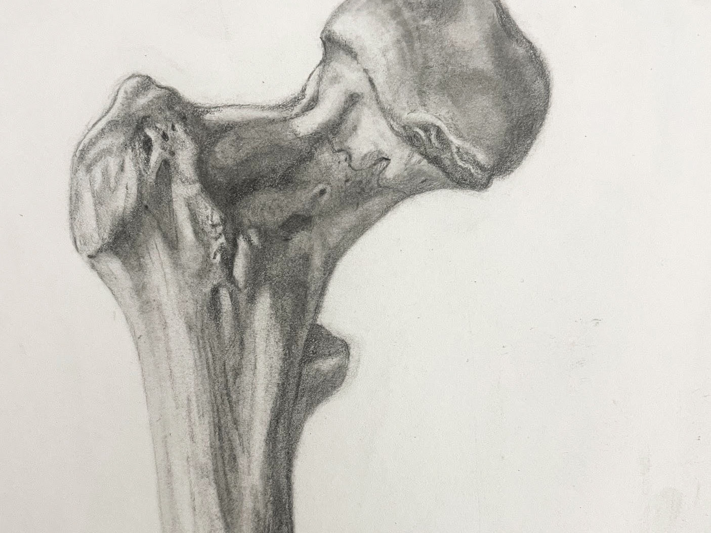
Initial Sketches:
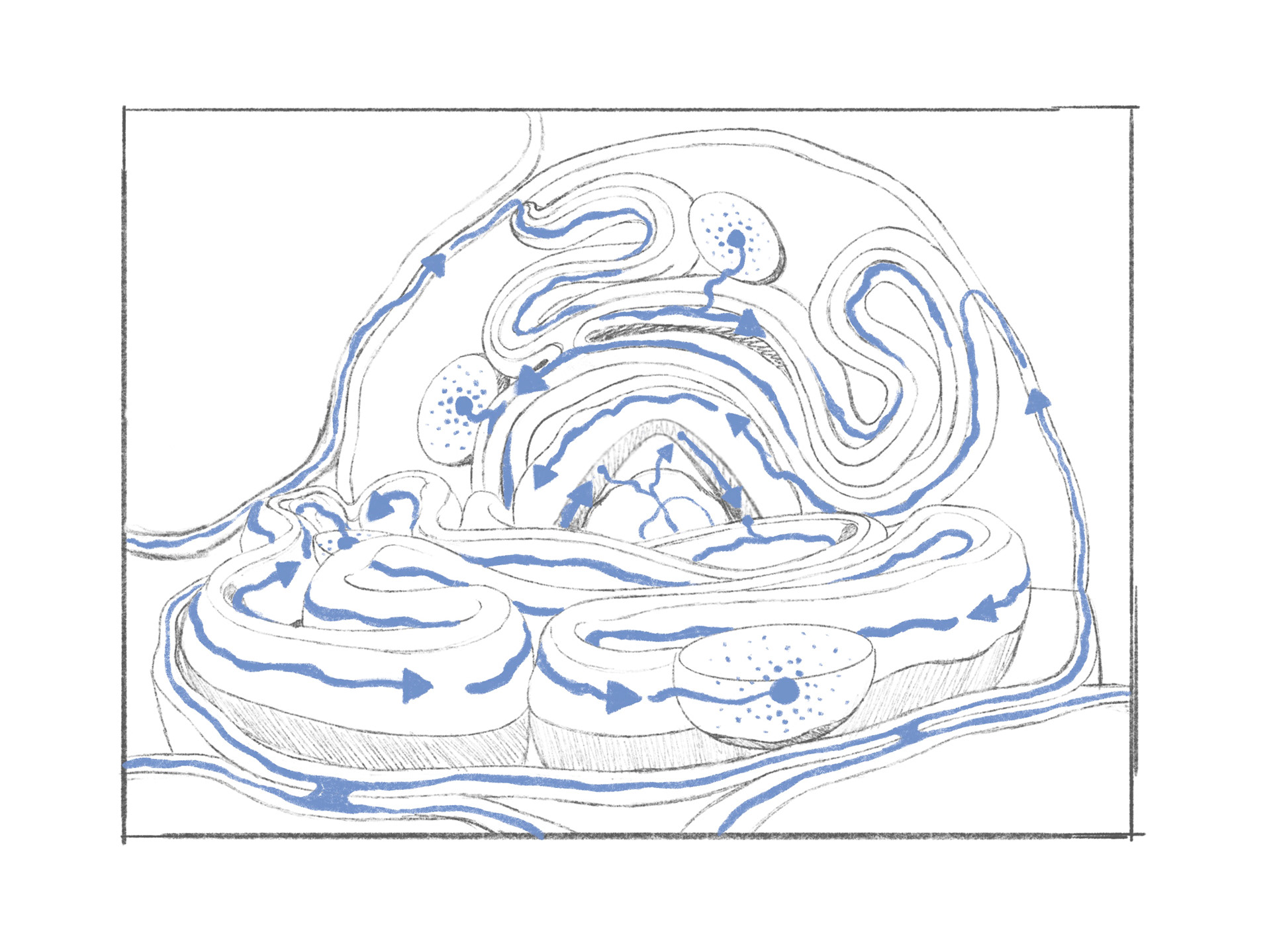
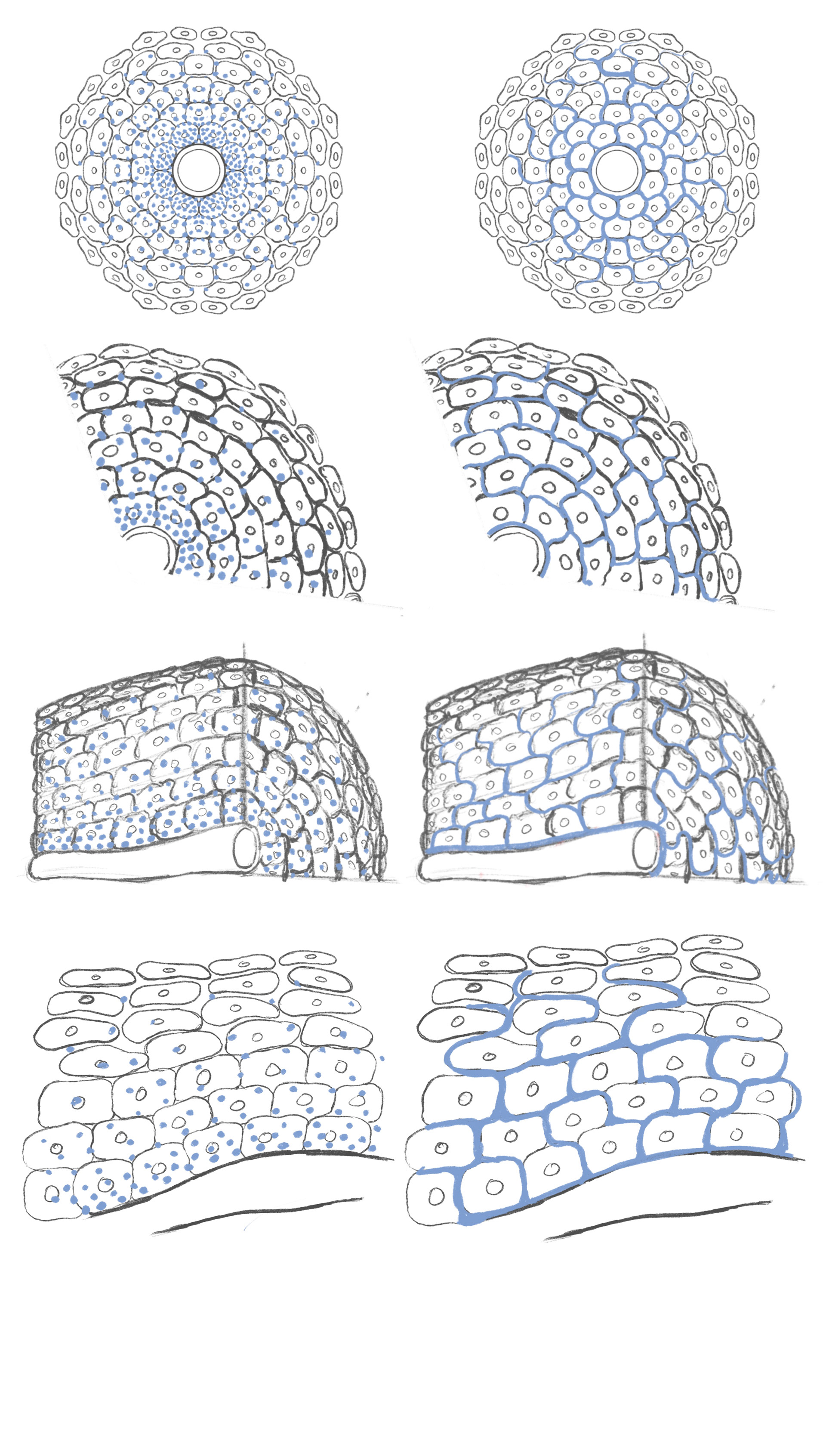
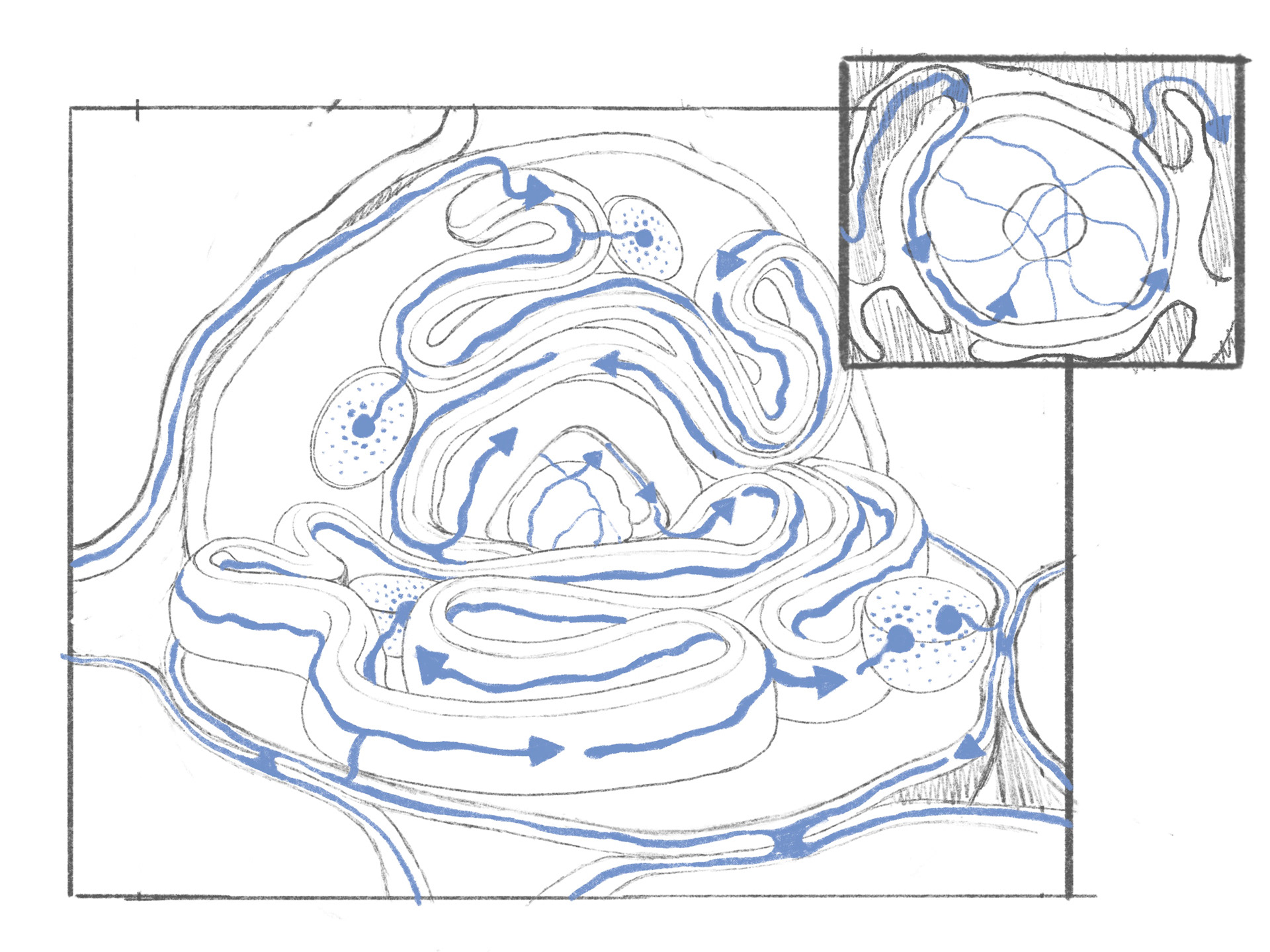
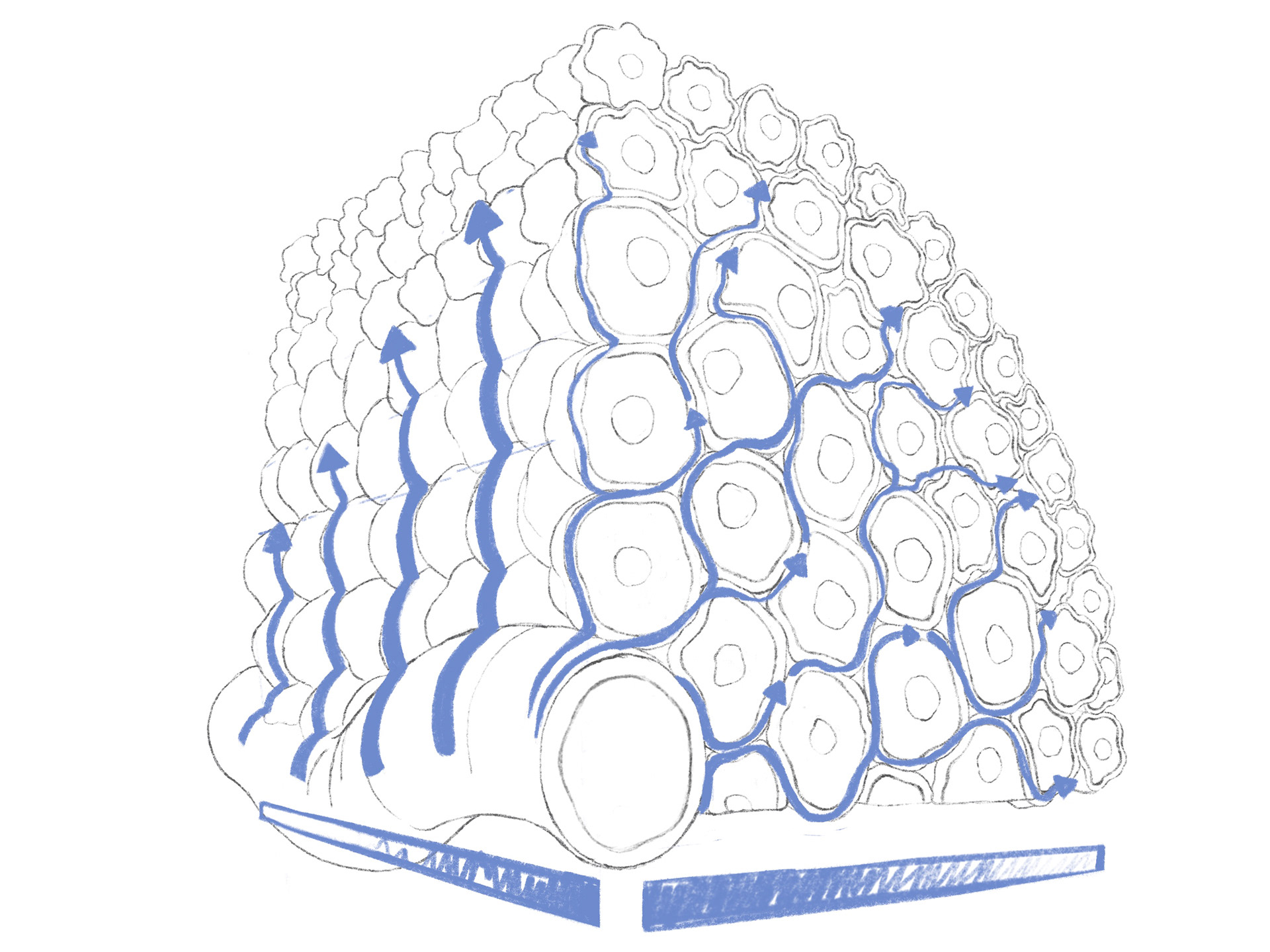
The process began with several exploratory sketches, each showcasing the concept in different visual ways. Through iterative feedback loops and close collaboration with the client, these initial ideas were refined and narrowed down to three final illustrations: one comparing the old and new models, and a detailed depiction of how oxygen moves through a single cell. These visuals were designed to bring clarity and impact to his academic paper.
3D Perspective References:
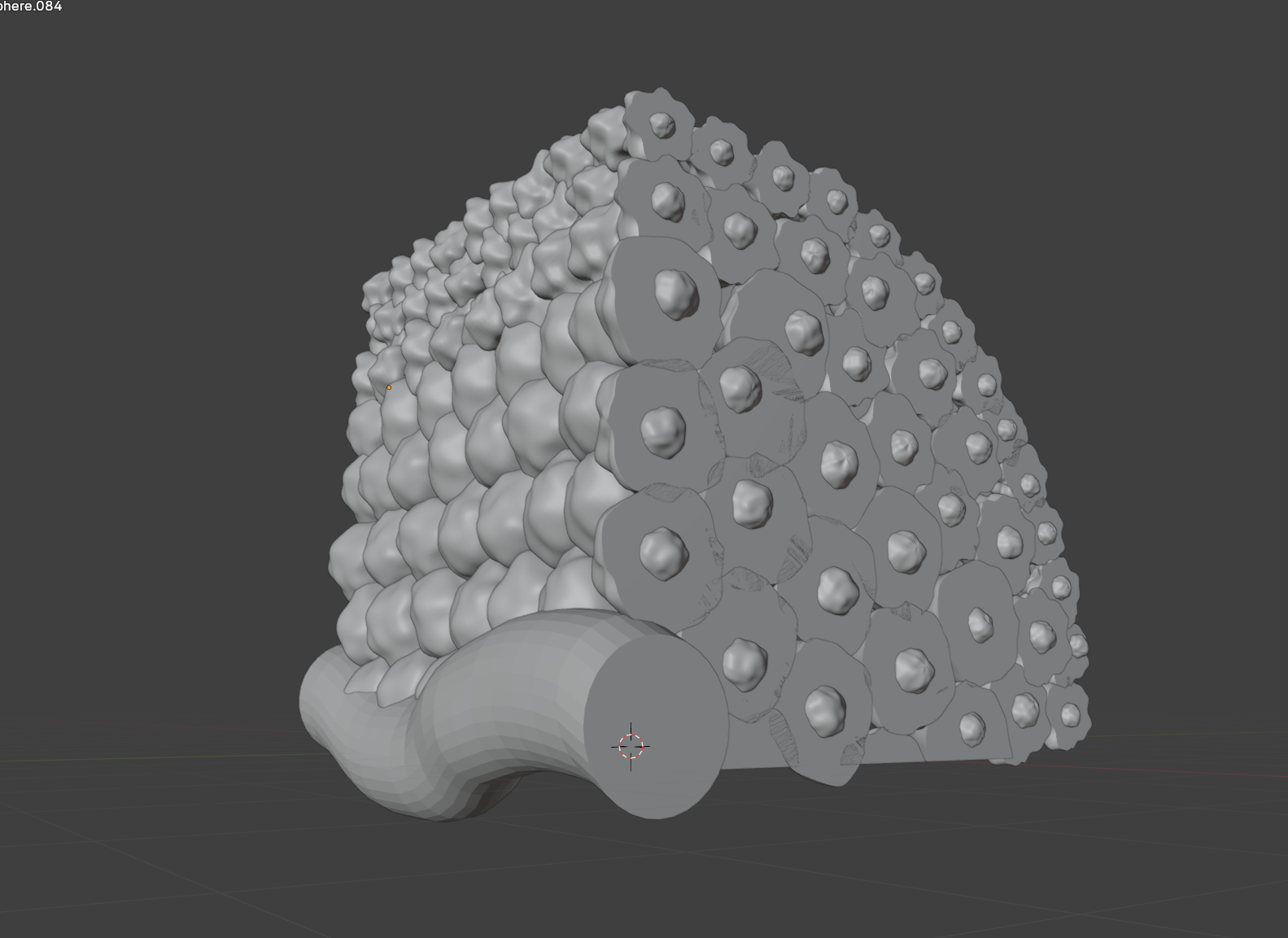
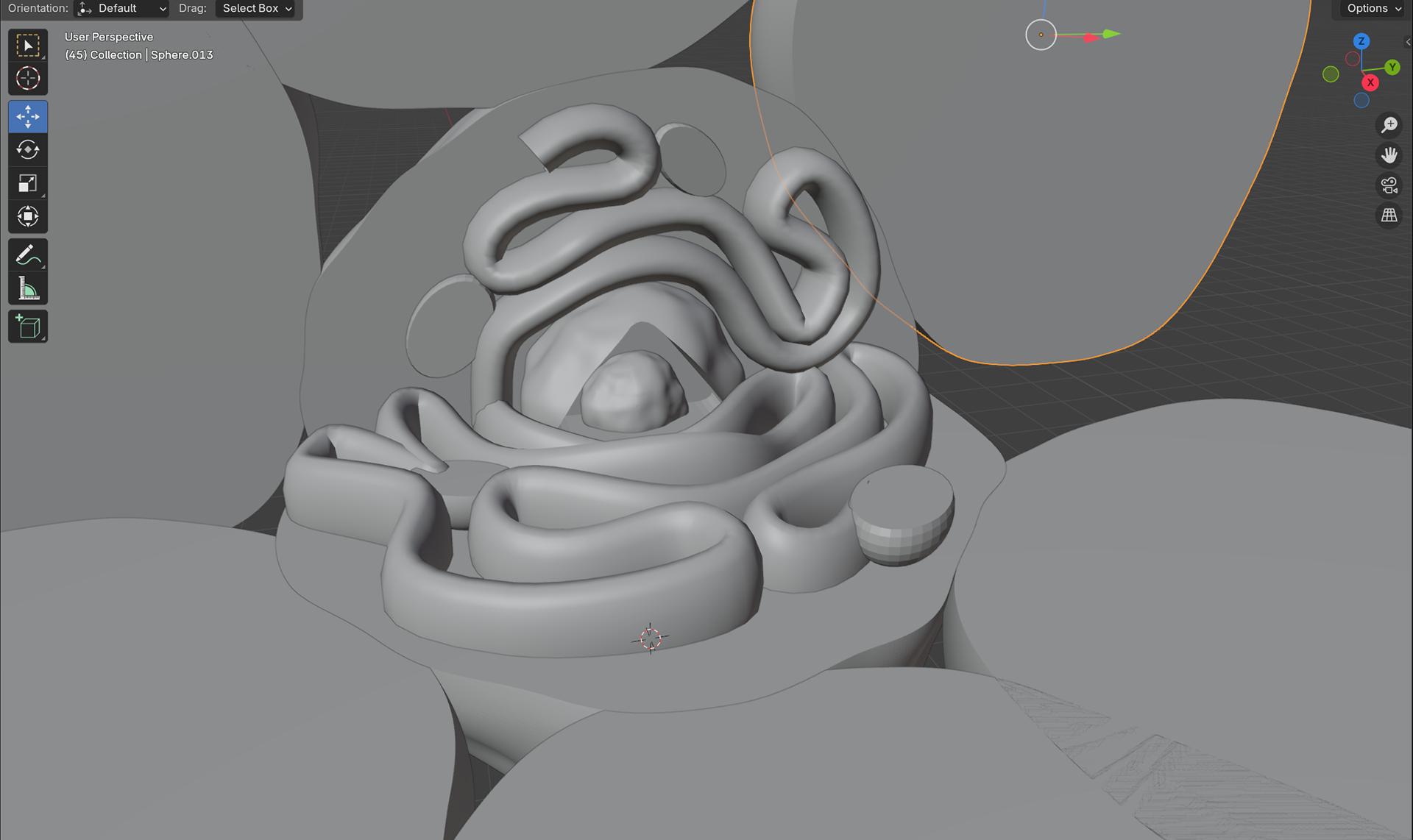
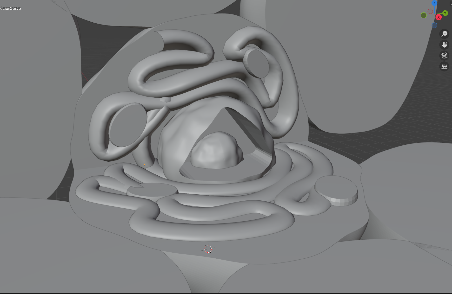
To ensure the perspective in my final illustrations was both accurate and realistic, I created perspective references in Blender using simple 3D shapes. By building basic forms that mirrored the structures in my sketches, I was able to experiment with camera angles, depth, and lighting to find the most effective composition. These 3D references served as a precise foundation for my final drawings, allowing me to maintain correct proportions and spatial relationships while still focusing on the illustrative and scientific details of the concept.


Cleaned Up Sketches:
After finalizing the foundational sketches, I refined the illustrations with a focus on guiding the viewer’s eye along the precise path that oxygen takes through the cells. Through multiple rounds of iterative feedback with my client, we experimented with visual techniques such as line weight, color , and compositional flow to emphasize the movement and directionality of the oxygen. This collaborative process ensured that the final images not only conveyed scientific accuracy but also directed attention seamlessly to the core concept of the new model.
Color Selection:
To ensure the illustrations were effective in both color and black-and-white formats, I experimented with various color schemes that maintained clarity and contrast when desaturated. After testing multiple approaches, I landed on a monochromatic orange-yellow palette, which provided warmth and depth to the cellular structures. I highlighted the oxygen pathways with light white and blue tones, creating a distinct visual contrast that allowed the oxygen to stand out prominently in both the full-color version and potential black-and-white reprints.

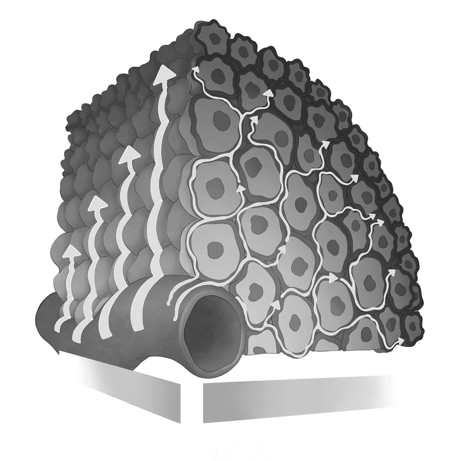
Black and White Vs Color Images: